Figure 3.—
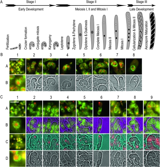
QIP is essential for the completion of early sexual development. (A) Sexual development in N. crassa. The cartoon diagram represents the three main stages (early development, middle development, and late development), from fertilization to ascospore maturation observed during sexual development. Drawings are not to scale. Cartoon was inspired on the work of Raju (1980). (B). Early wild-type development. Maternal and ascogenous tissue corresponding to a wild-type cross (DLNCR553 × DLNCR554; Table 2) was examined. In 1A, 1B, and 2A−8A, GLE-1–sGFP signal is green, whereas DAPI signal is red. 1A shows representative maternal tissue. 1B shows a 10× enlargement of one of the nuclei present in 1A. 2A−8A show merge images of developing croziers as they go through formation, conjugate mitosis, karyogamy, and early meiosis I. 2B−8B show light images. (C). Early QipΔ development. Maternal and ascogenous tissue corresponding to a mutant Qip loss-of-function cross (RMNCR113 × RMNCR114; Table 1) was examined. In 1A, 1B, 1C, and 1D and 2A, 2B−9A, 9B, GLE-1–sGFP signal is green. Whereas in 1A, 1B, 1C, and 1D and 2A, 2C−9A, 9C, DAPI signal is red. 1A and 1C show representative maternal tissue from days 3 and 5 postfertilization, respectively. 1B and 1D show a 10× enlargement of one of the nuclei present in 1A and 1C, respectively. 2A to 9A show merge images of developing croziers as they go through development. 2B to 9B show GLE-1–sGFP localization. 2C−9C show DAPI signal. 2D−9D show light images. All tissues were harvested at 3 and 4 days postfertilization, unless otherwise noted, and processed as described in materials and methods. Pictures were taken at 600×. The bar on 1A equals 20 μm.

