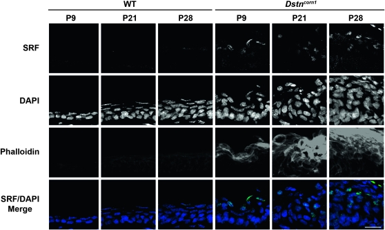
Figure 1.—
SRF expression and F-actin distribution in WT and Dstncorn1 cornea. Immunofluorescence shows that SRF (green) is detectable within the suprabasal nuclei of Dstncorn1 corneal epithelium as early as P9 and persists throughout the thickening of the epithelial layer and F-actin (phalloidin, red) accumulation. SRF is not detectable in WT cornea at any timepoint tested. All slides were counterstained with DAPI (blue). Bar, 20 μm.

