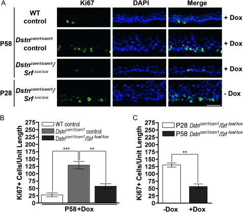
Figure 3.—
Corneal epithelial cell proliferation in WT control, Dstncorn1/corn1 control, and Dstncorn1/corn1/SrfΔce/Δce corneal epithelium. (A) Immunofluorescent staining for the cell proliferation marker Ki67 (green) demonstrates an obvious decrease in the number of proliferating cells in Dox-induced Dstncorn1/corn1/SrfΔce/Δce mice as compared to Dox-induced Dstncorn1/corn1 control mice. This is also decreased compared to Dstncorn1/corn1/SrfΔce/Δce mice prior to Dox treatment, demonstrating a reversal of the hyperproliferative condition. All slides were counterstained with DAPI (blue). Bar, 20 μm. (B) Quantification of the Ki67 positive cells throughout the length of the corneal epithelium confirmed a significantly smaller number of proliferating cells in Dox-induced Dstncorn1/corn1/SrfΔce/Δce mice as compared to Dox-induced Dstncorn1/corn1 control. (C) Quantification of the Ki67 positive cells throughout the length of the corneal epithelium confirmed a significant regression of the hyperproliferative phenotype following Dox induction in Dstncorn1/corn1/SrfΔce/Δce mice. Error bars, SEM. * denotes statistical significance. *P < 0.05, **P < 0.01, ***P < 0.001.

