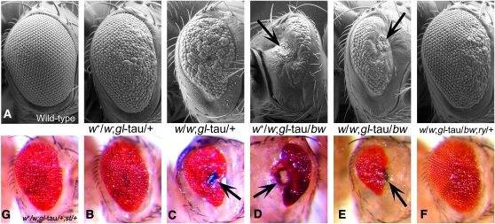
Figure 1.—
Scanning electron micrographs (SEMs) and color light micrographs demonstrating null alleles of white (w) and brown (bw) enhance tau-induced toxicity. Arrows: necrotic patches. (A) Wild-type (Canton-S). (B) white heterozygote: w+/w1118; gl-tau/+. (C) white homozygote: w1118; gl-tau/+. (D) brown allele: w+/w1118; bw1/gl-tau. (E) white homozygote + brown: w1118; bw1/gl-tau. (F) Null allele of rosy revert white and brown enhanced toxicity: w1118; bw1/gl-tau; ry506/+. (G) Null mutations in scarlet (st) do not affect tau-induced toxicity: w+/w1118; gl-tau/+; st1/+. Flies were anesthetized with carbon dioxide for light microscopy images, taken with a digital-camera equipped Zeiss dissecting microscope. Flies were dehydrated in hexamethyldisilazane prior to mounting for SEM, as previously described in Jackson et al. (2002). SEM images were taken on a Hitachi S-2460N scanning electron microscope. Stocks and crosses were maintained on a standard yeast-molasses-cornmeal medium at 23° or 25°.

