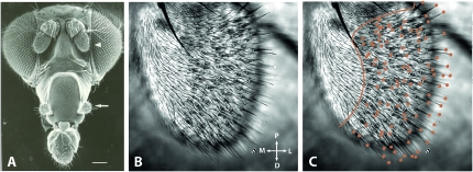
Figure 1.—
The olfactory organs and sensilla of D. melanogaster. (A) Electron micrograph of a Drosophila head indicating the two main olfactory organs, the antenna (arrowhead) and maxillary palp (arrow). Scale bar is 100 μm. (B and C) Transmitted-light confocal image showing the anterior face of a Drosophila third antennal segment. In C, the orange line separates the proximomedial region, which is devoid of trichoid sensilla, from the central and distolateral regions of the antenna, which are covered by basiconic, coeloconic, and trichoid sensilla. Orange dots indicate the tips of visible trichoid sensilla. Abbreviations: P, proximal; D, distal; M, medial; L, lateral. Image in A reproduced from Carlson(1996) with permission from Elsevier.

