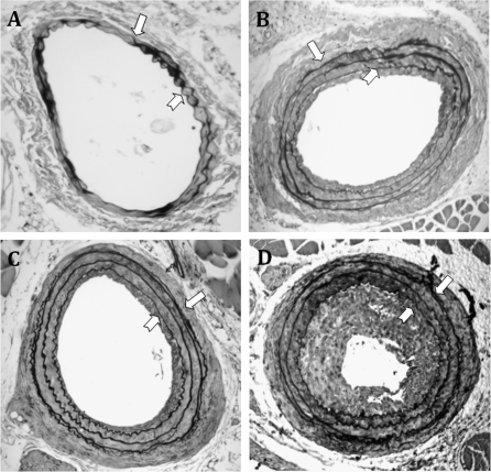
FIG. 4.
Representative Verhoeff's van Gieson staining of the injured carotid arteries from C57BL/6 mice treated with (A) control, (B) AZT, (C) indinavir (IDV), or (D) AZT + IDV. Digitalized images were acquired, and the neointimal and medial areas were analyzed as described in Materials and Methods. The block arrows indicate the EEL, and the notched arrows indicate IEL. The images were taken at a magnification of ×100.

