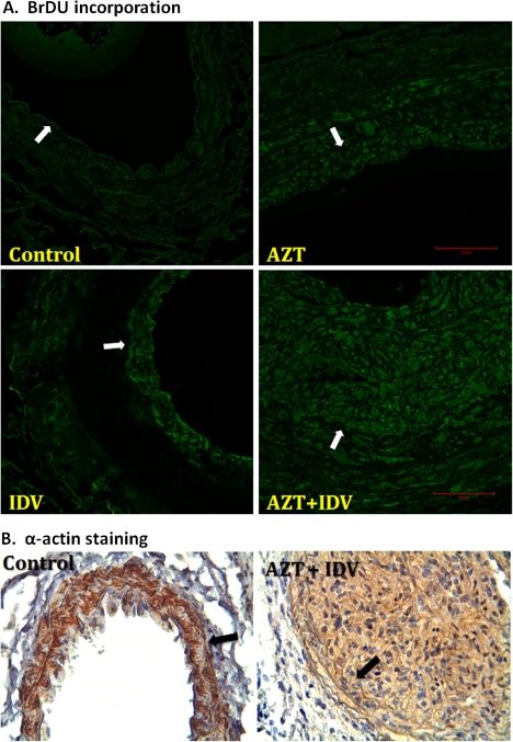
FIG. 6.
BrdU fluorescence (A) compared with smooth muscle α-actin staining (B) of proliferating cells in injured carotid arteries from C57BL/6 mice treated with antiretrovirals. Paraffin-embedded tissues were obtained 14 days after the endothelial injury procedure. For BrDU incorporation, injured artery cross-sections were incubated with an anti-BrdU primary antibody and then a secondary antibody conjugated to a fluorophore. The images were visualized at 488 nm. α-Actin was visualized using immunohistochemistry with hemotoxylin and eosin counterstaining. Brown staining thus indicates smooth muscle. The arrows denote IEL. The scale bar indicates 50 μm.

