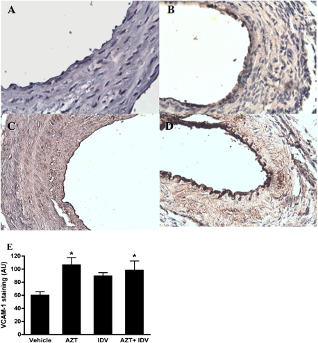
FIG. 8.
Representative immnuhistochemical staining for VCAM-1. Paraffin-embedded cross-sections from the animals that underwent the injury procedure were stained with a polyclonal antibody against mouse VCAM-1. Images represent immunostaining observed for the (A) control, (B) AZT-, (C) indinavir (IDV)-, (D) AZT + IDV–treated groups. Images were taken at a magnification of ×20. (E) Endothelial staining intensities for VCAM. Data are means ± SE. One-way ANOVA revealed a significant effect of treatment. Asterisk indicates p < 0.05.

