Fig. 4.
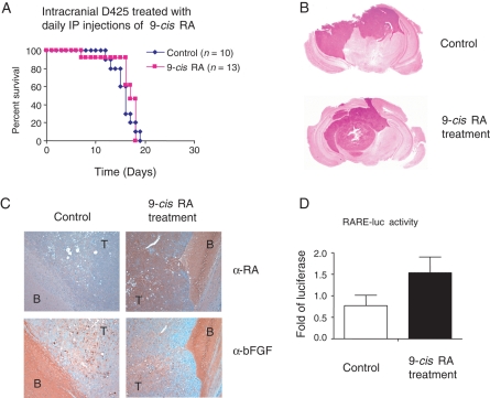
9-cis RA does not extend survival in mice with intracranial MB tumors. (A) No survival advantage in treatment by 9-cis RA. One million D425 cells were intracranially injected into mice and treated by IP injection of 50 µL of 9-cis RA dissolved in DMSO (n = 13) or treated with DMSO as control (n = 10). The daily treatment of the first 6 days was followed by a pause of 2 days and 5 additional daily injections. (B) Sections of H&E staining of mouse brains with or without the treatment by 9-cis RA. The darker nucleus-rich area represents the tumor formed by D425 cells. (C) α-RA and α-bFGF staining of mouse brains with or without the 9-cis RA treatment. Brain slides shown in (B) were stained with α-RA or α-bFGF and the positive staining was visualized by DAB colored in brown with hematoxylin in blue as counterstaining. Slides of serial cut of 10 µm apart were used and shown in ×10 magnification. Compared with the control, the brain with 9-cis RA treatment revealed an increased level of RA and generally unchanged bFGF expression. T, tumor; B, brain. (D) Luciferase reporter assay with the brain lysates. RARE-luc reporter construct with RA-responsive element were transfected in P19 cells and incubated with lysates of brain samples treated with or without 9-cis RA. The relative luciferase activity was graphed as the fold increase compared with the control. Three brain samples were used in each group.

