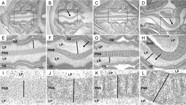
Figure 2.
Histological progression of PNS defects found in TEC1KO embryos. A–D, Magnified views (×4) of transverse sections of embryonic sinuses. For orientation, the brain is to the right in each panel, and the nasal openings are to the left. A, WT E14.5 embryo. Scale bar represents 500 μm and applies to images in panels A–D. B, E14.5 TEC1KO sinus showing slight deviation (arrow) of the PNS. C, WT E17.5 embryo. D, E17.5 TEC1KO embryo. Note that septal deviation is exacerbated at this stage (arrow). E–H, Magnified views (×10) of the boxed areas from panels A–D, respectively. E, WT E14.5 embryo. Note the thickness of the LP (bar). Scale bar represents 250 μm and applies to images in panels E–H. F, E14.5 TEC1KO sinus showing edema (arrow) and thickening (bar) of the LP. G, WT E17.5 embryo; the bar indicates thickness of the LP. H, Mutant E17.5 sinus showing increased edema (arrow) and thickening (bar) of the LP. I–L, Views (×40) of the PNS from panels E–H, respectively. The black bars represent thickness of the septum, which was significantly increased in mutants by E17.5 (panel L, P < 0.0001). Scale bar in panel I represents 50 μm and applies to images in panels I–L. OE, Olfactory epithelium.

