Figure 5.
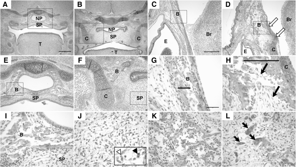
Histology of the E17.5 palate and craniofacial bones. A and B, View of coronal sections through the embryonic palate. A, WT embryo showing completely fused secondary palate (SP). Scale bar represents 500 μm and applies to images in panels A and B and E and F. Panel B, TEC1KO palate. Note that the secondary palate is fused; however, it is dramatically thicker than normal. Also note that the nasopharynx (NP) is collapsed due to loss of bony support. C and D, Views (×10) of the frontal bone. Note the presence of cartilage in the mutant (panel D, arrows) that is not found in the WT (panel C). Scale bar in panel C represents 250 μm and applies to images in panels C and D. Panels E and F, The boxed areas in panels A and B, respectively, are shown at ×10 magnification. Note the erroneous cartilage flanking the mutant (panel F). Also, note that the bone found in WT (panel E) encircling the nasopharynx is found only lateral to the palate in TEC1KO. G and H, Magnification (×40) of the areas boxed in panels C and D, respectively. Bone trabeculation (bars) was thicker and disorganized in mutant (panel H) compared with normal (panel G), and there was increased deposition of fibrous connective tissue (arrows). Scale bar in panel G represents 50 μm and applies to the images in panels G–L. Panels I and J, Magnified view of the boxed areas in panels E and F, respectively. Panel I, WT bone adjacent to the nasopharynx. Panel J, TEC1KO secondary palate. Note the absence of bone and the presence of apoptotic cells (inset, white arrowhead) and mitotic figures (inset, black arrowhead). Panels K and L, View (×40) of WT (panel K) and TEC1KO (panel L) mandible. Note the increased fibrous connective tissue and numerous osteoclasts (arrows) indicating osteolysis in the TEC1KO. B, Bone; Br, brain; C, cartilage; E, eye; T, tongue.

