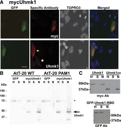
Figure 2.
Uhmk1 is present in both nucleus and cytosol. A, AtT-20 cells expressing mycUhmk1 and GFP were examined 24 h after transfection using antibody to myc or Uhmk1. Primary antibody was visualized using Cy3-tagged secondary antibody whereas nuclei were visualized using TOPRO3. Arrow marks moderate and asterisk marks low expressing cell; scale bar, 10μm. B, MycUhmk1 was transiently expressed in AtT-20 or PAM-1 AtT-20 cells; nuclei were isolated and 1% of the homogenate (H), supernatant (S), and nuclear (N) fractions was analyzed by SDS-PAGE; exogenous Uhmk1 was visualized using rabbit polyclonal antibody JH1998. *, Nonspecific band. C, AtT-20 PAM-1 cells were transfected with vectors encoding mycUhmk1, mycUhmk1cc, or GFP-Uhmk1-RBD. Cells were harvested the next day, and nuclei were isolated and analyzed. Antibodies used for Western blots are indicated below each blot. Ab, Antibody.

