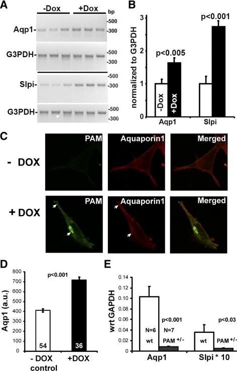
Figure 8.
Verification of array analysis. The cDNA prepared from noninduced and induced iPAM subclones was subjected to RT-PCR using PCR primers for aquaporin1 (Aqp1, NM_007472) and secretory leukocyte peptidase inhibitor (Slpi, NM_011414). G3pdh (NM_008084.2) transcript levels were evaluated simultaneously. A, Ethidium bromide-stained gels are shown. B, RT-PCR signals from Aqp1 or Slpi were normalized to G3PDH for each subclone. Data are mean ± sd; P values calculated using Excel (t test, unequal variances). C, iPAM cells grown in the absence or presence of doxycycline (72 h) were stained simultaneously for PAM-1 (green) and Aqp1 (red). Arrows mark colocalization and accumulation of PAM-1 and Aqp1 at the TGN and at the distal tips of processes, where LDCVs accumulate. D, Aqp1 staining intensity in iPAM cells grown in the absence (n = 54) or presence (n = 36) of doxycycline (as in panel C) was quantified; data are mean ± sem (t test, unequal variance). E, The expression of Aqp1 and Slpi was analyzed by quantitative PCR using pituitary RNA from wild-type control mice (wt; n = 5) and PAM heterozygote mice (PAM+/−; n = 6); G3PDH was analyzed simultaneously, and data were calculated relative to G3PDH (GAPDH) for each sample using the ΔCT method (65); data are mean ± sem. Dox, Doxycycline.

