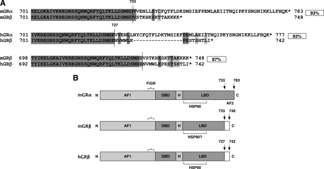
Figure 3.
Sequence and functional domain comparisons of mGRβ and hGRβ. A, C-terminal regions of GRα and GRβ were aligned for each species. Overall percent homologies for entire proteins are indicated. Vertical lines indicate borders between exon 8 and distal domains. Light gray boxes indicate conservative substitutions. B, The mGRβ isoform exhibits a functional domain structure that is nearly identical to hGRβ. Compared with mGRα, the β-isoforms of both species have reduced and distinct C-terminal regions that lack the activation function-2 (AF-2) domain (helix 12). These features account for their reduced ability to bind hormone and activate transcription. DBD, DNA-binding domain; H, hinge region; LBD, ligand-binding domain; FiGR, epitope recognized by FiGR monoclonal antibody. HSP90 binding regions of mGRα and hGRβ are shown, along with putative site in mGRβ.

