Figure 2.
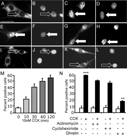
CCK induces EGR1 translocation to the nucleus in cultured vagal afferent neurons, and this does not depend on EGR1 expression. Vagal afferent neurons cultured for 72 h and stained with antibody to EGR1 (A, C, E, G, I, and K) and DAPI (B, D, F, H, J, and L) in respective fields. A and B, Nuclear EGR1 staining was undetectable after transfer to serum-free medium for 2 h; C and D, nuclear EGR1 was readily detectable after treatment with 10 nm CCK8s (1 h) in serum-free medium; E–H, the effect of CCK was not inhibited by pretreatment with the transcriptional inhibitor actinomycin D (2 μm) (E and F) or the translational inhibitor cycloheximide (10 μg/ml) (G and H); I–L, nuclear EGR1 was undetectable after treatment with ghrelin (10 nm) (I and J), but ghrelin (10 nm) inhibited the effect of 10 nm CCK8s on nuclear localization of EGR1 (K and L). Arrows identify a representative neuron in each pair of images: filled arrows show EGR1 localization to the nucleus, and open arrows show EGR1 excluded from the nucleus. M, Time course of CCK8s-induced nuclear translocation of EGR1; N, quantification of immunohistochemical data described above represented as the percentage of total cells exhibiting nuclear EGR1. ***, P < 0.001 compared with control; **, P < 0.01 compared with response to CCK8s. Representative images from six independent experiments are shown. Scale bar, 50 μm (A–L).

