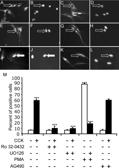
Figure 3.
CCK induces EGR1 localization to the nucleus through activation of PKC and Erk1/2 in vagal afferent neurons. Vagal afferent neurons cultured for 72 h and stained with antibody to EGR1 (A, C, E, G, I, and K) and DAPI (B, D, F, H, J, and L) in respective fields (see Fig. 2 for further details). A–D, The PKC inhibitor Ro32-0432 (1 μm) on its own had no effect on EGR1 localization (A and B) but inhibited the action of 10 nm CCK8s (1 h) on nuclear localization of EGR1 (C and D); E–H, the inhibitor of Erk1/2 activation U0126 (10 μm) had no effect on EGR1 translocation on its own (E and F) but inhibited the effect of 10 nm CCK8s (G and H); I–L, nuclear EGR1 localization was readily detectable after treatment with 100 nm PMA (1 h) (I and J), and this effect was inhibited by 10 μm U0126 (K and L). In A–L, arrows identify a representative neuron in each pair of images: filled arrows show EGR1 nuclear localization, and open arrows show EGR1 exclusion from the nucleus. M, Quantification of data in A–L represented as the percentage of total cells exhibiting nuclear EGR1 and response to CCK8s (10 nm) in the presence of AG490 (50 nm), an inhibitor of STAT3. ***, P < 0.001 compared with the relevant control. Representative images from six independent experiments are shown. Scale bar, 50 μm (A–L).

