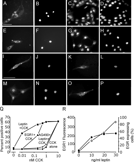
Figure 4.
The action of CCK on nuclear translocation of EGR1 is potentiated by overexpression of EGR1 and by leptin. Vagal afferent neurons cultured for 72 h and stained with antibody to EGR-1 (A, C, E, G, I, K, M, and O) and DAPI (B, D, F, H, J, L, N, and P) in respective fields. A and B, Transfection with an EGR1 overexpression vector was associated with EGR1 immunoreactivity in the cytoplasm but not nuclear translocation; C and D, addition of 10 nm CCK8s induced EGR1 localization to the nucleus in overexpressing neurons; E and F, neurons stimulated with leptin (30 ng/ml) exhibited EGR1 localization to the cytoplasm, but not nucleus; G and H, stimulation with both leptin (30 ng/ml) and CCK8s (10 nm) led to nuclear EGR1; I and J, with lower concentrations of leptin (10 ng/ml), there was again EGR1 in the cytoplasm; K and L, low doses of CCK8s (1 nm) had no effect of EGR1 translocation; M and N, importantly, 1000-fold lower concentrations of CCK8s (10 pm) in the presence of 10 ng/ml leptin induced the nuclear translocation of EGR1; O and P, there was no effect of 10 ng/ml leptin on EGR1 nuclear translocation in cells stimulated with very low concentrations of CCK8s (2 pm); Q, quantification of cells exhibiting nuclear EGR1 staining in response to varying concentrations of CCK8s alone; CCK8s and leptin (10 ng/ml); CCK8s, leptin (10 ng/ml), and AG490 (50 nm); or CCK8s and EGR1 overexpression; R, the intensity of EGR1 fluorescence in vagal afferent neurons is shown in response to leptin: left, mean fluorescent intensity (○) in cells where fluorescence was more than 2 sd above mean of unstimulated cells; right, numbers of cells (•) with fluorescent intensity more than 2 sd above the mean of unstimulated cells (n = 6).

