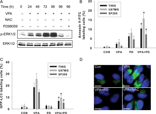
Fig. 6.
Role of the ERK pathway in VPA-induced autophagy. (A) Representative Western blot showing ERK 1/2 phosphorylation in U87MG cells after treatment without (control) or with VPA (1.0 mM) alone for 24–96 h or with PD98059 (10 µM) or NAC (5 mM). (B) Apoptosis measured using annexin V-FITC staining. Cells were treated with 1.0 mM VPA alone or with 10 µM PD98059 for 96 h. Data were the mean of triplicate experiments; error bars, SD *P < .05, as compared to the VPA-treated group. (C) Quantitation of cells with GFP-LC3 dots among cells treated with 1.0 mM VPA alone or 1.0 mM VPA and 10 µM PD98059. Cells were transiently transfected with the GFP-LC3 plasmid for 24 h and then treated for 96 h as described. The number of cells with GFP-LC3 dots was counted, and their percentage among the total number of cells expressing GFP was determined. Data were the mean of triplicate experiments; error bars, SD *P < .05, when compared with the VPA-treated group. (D) Representative immunostaining of p-ERK1/2 in U87MG cells with VPA (1.0 mM) alone or with PD98059 (10 µM) and NAC (5 mM) for 96 h. Bar, 10 µm.

