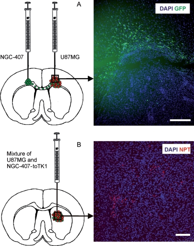
Fig. 2.
(A) NGC-407 cells display tropism toward xenografted GBM. U87MG GBM cells were injected into the right hemisphere and NGC-407-GFP cells into the left hemisphere, both at the level of the corpus callosum. The cartoon shows the green NGC-407 cells migrating through the corpus callosum toward the GBM xenograft. Immunohistochemistry showing GFP-positive NGC-407 cells infiltrating the tumor bed. High cell density identified by DAPI staining localizes the tumor area. Scale bar is 100 µM. (B) NGC-407-toTK1 cells stained red with NPT II are dispersed throughout the U87MG xenograft and intermingling with the tumor cells. The cartoon is showing the coimplantation of NGC-407-toTK1 and U87MG cells into the right hemisphere. NPT was introduced into the NGC-407 cells for selection purposes during the myc immortalization phase. Scale bar is 100 µM.

