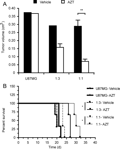
Fig. 4.
NGC-407 NPCs expressing toTK1 reduce tumor volume and improve survival in animals with human GBM xenografts. NGC-407 cells expressing toTK1 were coimplanted with U87MG glioma cells at 1:3 (n = 6) or 1:1 (n = 6) ratios. Each group of animals was then subgrouped and treated with AZT or vehicle for up to 21 days starting 24 hours after implantation. U87MG cells alone were injected in another group (n = 6), which was also treated similarly. (A) Tumor volumes after the stem cell delivery of toTK1 into intracranial GBM xenografts. Bar graphs show the volume of xenografts measured from multislice MRI images in the sagittal, coronal, and axial planes on day 22 or 23 postimplantation. Comparisons were made between AZT and vehicle treatment. **P < .01 (2-tailed, α = 0.05, n = 3; Student's t-test for unpaired values). Values are means ± SEM in the presence of error bars. (B) Kaplan–Meier survival plots showing improved survival on exposure to AZT. Comparison was made between these 2 treatment groups. *P < .05 (α = 0.05, n = 3) in both comparisons (log-rank [Mantel–Cox] test). The survival curves along with the curves for 1:30 ratio presented in Fig. 5 show a significant trend (P < .001, α = 0.05, n = 8; log-rank test for trend).

