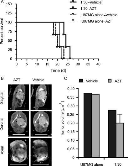
Fig. 5.
Effects of toTK1-expressing NGC-407 cells coimplanted with U87MG cells at a 1: 30 ratio. (A) Kaplan–Meier survival plots of nude rats that received only U87MG cells (n = 6) or the NGC-407 and U87MG cells (1:30; n = 6) and were exposed to AZT or vehicle for 14 days. P = .0629 for 1:30 group (α = 0.05, n = 3) and P = .4855 for U87MG-alone group (α = 0.05, n = 3), when treatment subgroups were compared (log-rank [Mantel–Cox] test). (B) Sagittal, coronal, and axial MRI images after coimplantation of NGC-407 and U87MG cells at a 1:30 ratio. One AZT-treated and 1 vehicle-treated animal are shown. MRI experiments were performed on day 22 or 23 postimplantation. Scale bar is 1 cm. (C) Bar graphs showing the volume of xenograft tumors after implantation of NGC-407 and U87MG cells at a 1:30 ratio or U87MG cells alone and upon exposure to AZT or vehicle. Volumes are measured from multislice images of MRI experiments.

