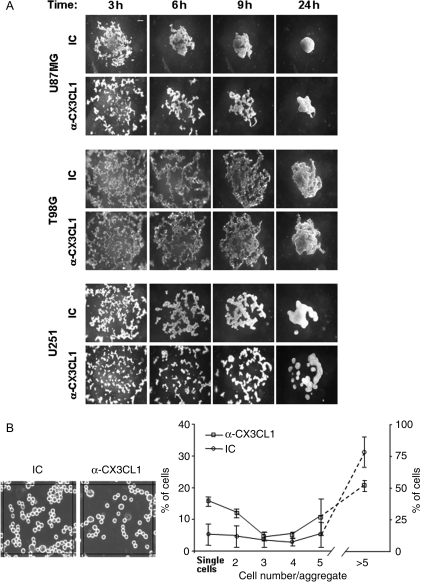
Fig. 4.
CX3CL1 is involved in glioma cell aggregation. (A) Slow aggregation assay on agar substrate was performed in the presence of 40 µg/mL of anti-CX3CL1 or IC mAb in 96-well plates, at 37°C, 5% CO2 as described in material and methods. Images shown were collected at 40× magnification and indicate a representative experiment of 3 independent experiments performed. Scale bar: 100 µm (B) Aggregation of T98G cells after 1 hour incubation in presence of 40 µg/mL of anti-CX3CL1 or IC mAb. Shown is a representative field (25 000 pixel2) obtained using Image J software. Data were expressed as the percentage of the mean ± SD of the number of single cells or aggregates containing 2 or more cells relative to a total of 500 cells, counted in several central fields.

