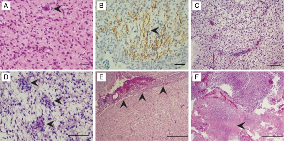
Fig. 3.
Putative aggressive pathological features in pilocytic astrocytoma specimens. (A–F) Putative aggressive features noted on review of 20 anonymized pilocytic astrocytoma specimens. Tumor infiltration demonstrated by entrapped neuronal cell bodies (arrowhead in A) and axons coursing through the neoplasm (arrowhead in B, neurofilament stain), oligodendroglioma-like features (C), pilomyxoid features (arrowheads in D), leptomeningeal spread (arrowheads in E), and necrosis (arrowhead in F) were noted in 9 of the patient samples. These pathological features were more prevalent in patients diagnosed prior to the age of 10 (Table 1). Scale bar indicates 50 µm in A and B, 100 µm in C and D, and 500 µm in E and F.

