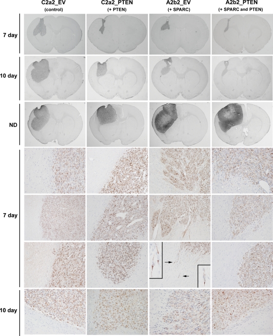
Fig. 7.
PTEN reconstitution suppresses the in vivo invasion of SPARC-expressing cells. Cells (400 000 in 5 µL) were intracranially implanted into nude rat brains and allowed to grow for 7 and 10 days, or until signs of neurological deficit. Brains were harvested, formalin-fixed, and paraffin-embedded. Tumor sections were stained for human mitochondrial marker to identify the tumor cells. Whole brain sections illustrating the largest cross sections through the tumors were captured at ×0.5 magnification. Days 7 and 10 sections were captured at ×20 magnification. Arrows indicate the single cell infiltration illustrated in the insets captured at ×60 magnification. Representative images of n = 6 (day 7) and n = 3 (day 10; ND) animals/group.

