Fig. 5.
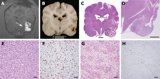
Necropsy data from Patient 1. Real-time imaging of infusions and availability of necropsy in all clinical cases allowed histopathological data to be correlated with areas of infused and noninfused tumor and normal brain. (A) T1-weighted real-time imaging of the final CED showing poor infusion of tumor tissue medially (arrow). (B) Gross pathological specimen at the same level as the MRI (scale bar = 1 cm). (C) Whole brain section (hematoxylin and eosin) showing distinct areas consisting of infused tumor with malacia (M), infused tumor with modified tumor (I), and noninfused tumor (T) (scale bar = 500 µm). (D) Magnified view of infusion area (hematoxylin and eosin, scale bar = 500 µm). Modification of tumor phenotype was seen in areas of tumor that were infused (G, hematoxylin and eosin) compared with noninfused tumor (E, hematoxylin and eosin). Infused tumor was less cellular with a more homogenous cellular phenotype. MIB-1 index in infused tumor (H) was <1% compared with 15% in noninfused tumor (E) (E, F, G, H, sacle bar = 60 µm).

