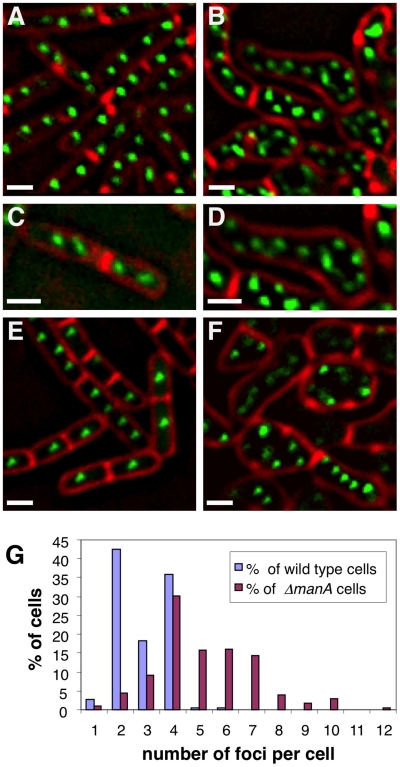
Figure 2. ΔmanA cells contain multiple copies of fully replicated chromosomes.
Origin and terminus regions were visualized in wild type and ΔmanA cells grown in rich LB medium. (A–B) Fluorescence images of typical wild type (SB294) (A) and ΔmanA (ME34) (B) cells producing Spo0J-GFP (green). Cells were stained with the membrane dye FM4-64 (red). (C–D) Enlargements of fluorescence images of wild type (C) and ΔmanA (D) cells as in (A–B). (E–F) Fluorescence images of wild type (ME79) (E) and ΔmanA (ME82) (F) cells bearing repeated tetO units inserted in proximity to the terminus region and producing TetR-GFP (green). Cells were stained with the membrane dye FM4-64 (red). (G) Quantification of Spo0J-GFP foci per cell in wild type (SB294) and ΔmanA (ME34) cells. At least 230 cells were analyzed for each strain. Scale bars correspond to 1 µm.

