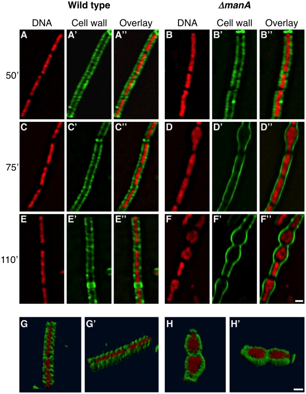
Figure 5. Following cell wall architecture and chromosome morphology.
Wild type (PY79) and ΔmanA (ME37) cells were grown in LB at 23°C and then shifted to 37°C. Samples were imaged at the indicated time points after temperature shift. (A–F) Superimposed fluorescence images of wild type (A, C, E) and ΔmanA (B, D, F) cells labeled with WGA-FITC (green) and stained with DAPI (red). (G–H) 3D reconstruction (see Materials and Methods) of wild type (PY79) (G,G') and ΔmanA (ME37) (H, H') cells labeled with WGA-FITC (green) and stained with DAPI (red) (see corresponding Videos S1 and S2, respectively). Scale bars correspond to 1 µm.

