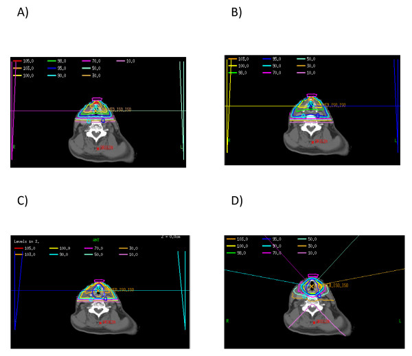
Figure 3.
Representative Axial Slices of Four Different Plans, a) tight clinical 2D plan, b) loose clinical 2D plan, c) 3D plan with 0.5 cm expansion from CTV to PTV, and d) IMRT plan with 0.5 cm expansion from CTV to PTV. The PTV is delineated in Figures 3c and 3d by the dark blue thick line encompassing the larynx.

