Figure 2.

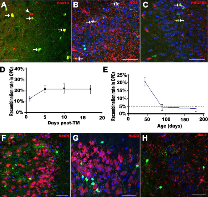
Genetic labeling of adult OPCs in the piriform cortex. A, Almost all EYFP+ cells expressed Sox10 (red). B, C, A subpopulation of EYFP+ cells were NG2+ (red) or PDGFRα+ (red) putative OPCs, respectively. D, Recombination rate in NG2+/PDGFRα+ OPCs on different days after last tamoxifen injection. Note that recombination reached a plateau after 5 d after TM. E, Recombination rates in NG2+/PDGFRα+ OPCs at different ages. Virtually no NeuN+ (red) (F) or HuCD+ (red) (G) cells were labeled before 17 d after TM. H, No Iba-1+ (red) microglia were labeled with EYFP. Arrows in A–C point to examples of double-labeled cells. Arrowhead in A, single-labeled cell. Scale bars, 50 μm.
