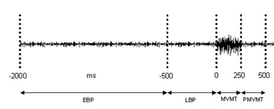
Figure 1.

Timing epochs used to capture SEPs relative to movement. Example of raw EMG from flexor digitorum superficialis of the hand performing the voluntary squeeze. Timing windows used to divide median nerve stimulations into respective epochs relative to the onset (0 ms) of EMG are shown. (EBP) Early Bereitschaftspotential (-2000 ms to -500 ms); (LBP) Late Bereitschaftspotential (-500 ms to -1 ms); (MVMT) Movement (0 ms to +250 ms); (PMVMT) Post-Movement (+251 ms to +500 ms).
