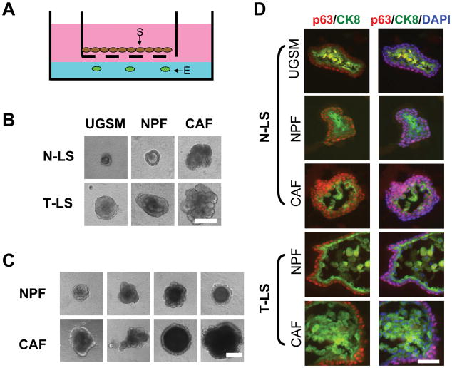
Figure 1. Analyses of spheroids formed from putative normal and cancer stem cells.
A. Illustration of the placement of an insert (pore size: 8.0 μm) containing stromal cells on top of the Matrigel for the co-culture experiments (E: epithelial cells; S: stromal cells). B. Phase contrast images of representative spheroids formed from either N-LS or T-LS cells after 14 days of co-culture with primary stromal cells from different sources. Bar, 100 μm. C. Illustration of the different sizes of the spheroids formed from tumor LSC cells after co-culturing with either NPFs or CAFs. Bar, 100 μm. D. Comparative immunohistochemical analysis of spheroids formed from normal and tumor LS cells co-cultured with UGSM, NPFs or CAFs. Sections of spheroids were analyzed by co-immunofluorescence using antibodies against the basal cell marker p63 (red) and luminal cell marker CK8 (green). DAPI was used for labeling cell nuclei. Bar, 25 μm.

