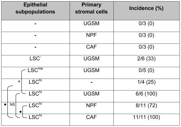
Table 1.
Detection of prostatic glandular structures in grafts.
 |
Grafts containing tumor epithelial subpopulation (104) and a type of primary stromal cells (104) were transplanted under kidney capsules. After 10 weeks, each animal was sacrificed, the kidney with the graft isolated, fixed, and then thin tissue sections were stained to determine the presence or absence of microscopically detectable glandular structures in the grafts. Statistical evaluation of the difference in incidence of detection of glandular structures between a marked pair of individual groups is indicated by
for p<0.05,
for p<0.01, or NS, for not significant.
