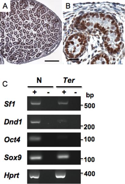
Fig.2. Expression of Sf1 in the testes.

(A) Immunostaining of PN1 testes from wild-type mice using anti-SF1 antibody at low (bar represents 200 um) and (B) higher magnifications (bar represents 20 um). (C) RT-PCR for Sf1, Dnd1, Oct4, Sox9 and Hprt using total RNA from PN1 testes of wild-type (N) and B6-Ter/Ter (Ter) mice. PCR was performed on equal amounts of cDNA. + indicates presence of Superscript during cDNA preparation. - are control lanes and indicates no Superscript was added.
