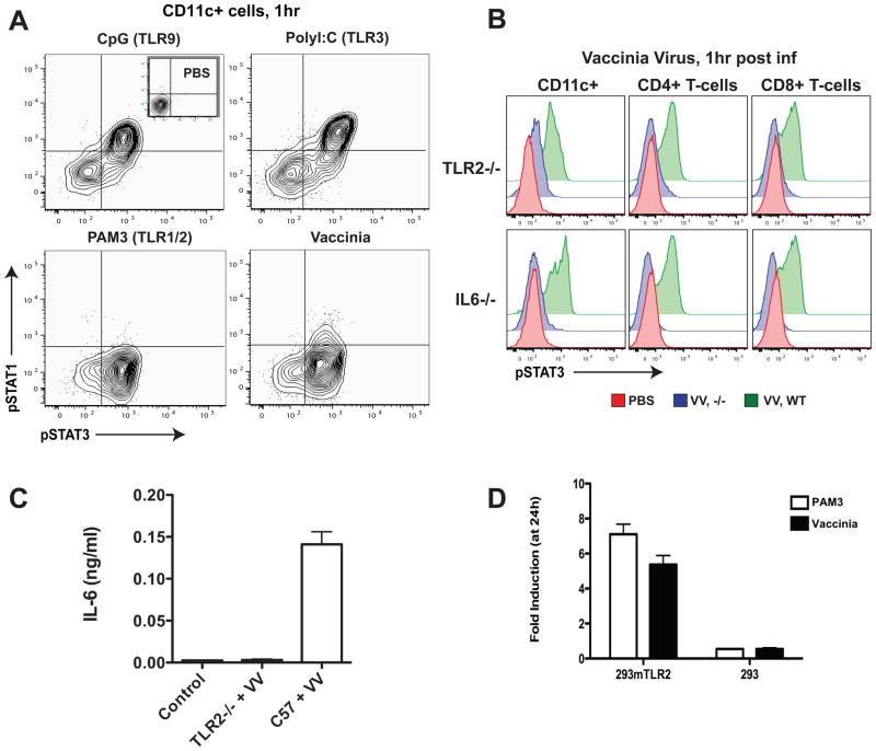
Figure 2.
Systemic vaccinia infection elicits TLR2 and IL-6-dependent responses that promote viral clearance and neutralizing antibody production.
(A) Early in vivo STAT response to vaccinia virus infection resembles the PAM3CSK4 (TLR1/2 ligand) response. pSTAT1 and pSTAT3 activation in CD11c+ cells at 1 hour after intravenous (i.v.) injection with PBS control, CpG, poly (I:C), or PAM3CSK4, compared to vaccinia virus infection (1 × 10⁷ pfu; Western Reserve strain).
(B) Vaccinia induction of pSTAT3 is TLR2 and IL-6 dependent. Wild-type (green), TLR2−/−, or IL6−/− (both in blue) mice were infected with vaccinia virus (VV) as before, and, after one hour, spleens were excised, dissociated, and prepared for intracellular analysis. pSTAT3 levels were determined in CD11c+, CD4+, and CD8+ cells.
(C) IL-6 production in response to vaccinia infection is TLR2 dependent. Wild-type and TLR2−/− mice were infected i.v. with 1 × 10⁷ pfu of vaccinia virus; after 1 hour serum was harvested for detection of secreted IL-6 by ELISA. Serum from uninfected mice was used as control.
(D) Vaccinia is recognized by TLR2 in vitro. HEK-293 cells transfected with mouse TLR2 and an NFκ B-driven luciferase reporter and untransfected control cells with the NFκ B-driven luciferase reporter alone were exposed to UV-inactivated vaccinia virus (5 viral particles per cell). NFkB-driven luciferase expression was evaluated by bioluminescence signal (after addition of luciferase) 24 hours later (data represented as mean ± SD).

