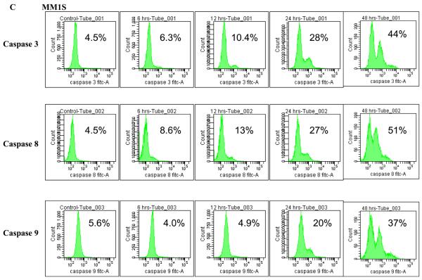
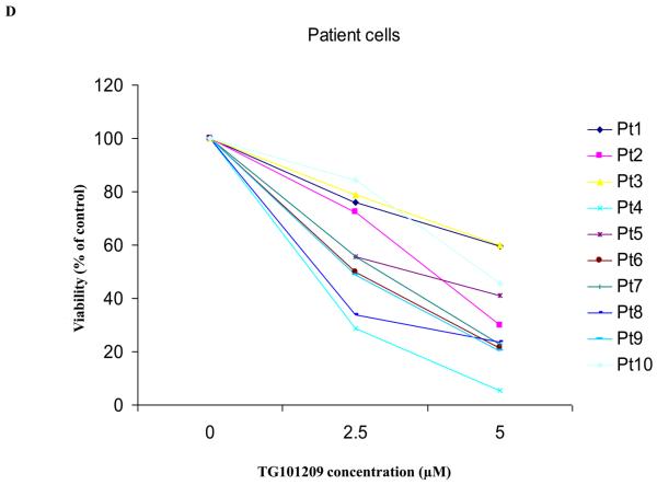
Figure 2. TG101209 induces apoptosis in MM cell lines and patient samples.
A) When MM1S or B) RPMI 8226 cells were incubated with TG101209 (5μM) we observed time dependent increase in apoptosis. Annexin V-FITC staining is represented on the X-axis and propidium iodide (PI) staining is represented on the Y-axis. Percent cells in viable quadrant are indicated. C) When MM1.S MM cells were treated with TG101209 (5 μM), induction of apoptosis in MM cells was accompanied by a time-dependent (0, 6, 12, 24 and 48 hours) cleavage of caspase 3, caspase 8, and caspase 9 as demonstrated by flow cytometry. Annexin V-FITC staining is represented on the X-axis and propidium iodide (PI) staining is represented on the Y-axis. Percent cells in viable quadrant are indicated. D) TG101209 induces apoptosis of freshly isolated patient MM cells when cultured with the indicated drug concentrations for 48 hours as measured using Annexin/PI staining and flow cytometry. TG101209 concentrations (μM) are indicated on the X-axis and % of viable cells (annexin and PI negative) is indicated on the Y-axis.


