
Figure 7.
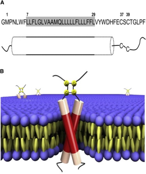
(A) Proposed model of the membrane-inserted structure of E5. The gray box represents the putative transmembrane segment that forms the regular helix fraction (solid outline), which is flanked by distorted helical regions (dotted outline) that extend beyond the lipid bilayer. The cysteines are located in the adjacent unstructured stretch near the C-terminus. (B) Three-dimensional scheme of the E5 dimer in a lipid bilayer based on the model in A. The regular helix parts are shown as red and the distorted helical regions as pale red in the cylinders, and the unstructured parts are shown as black lines. The cysteines are arranged to allow parallel disulfide bridges between Cys37-Cys37 and Cys39-Cys39. The sketch was made using POV-Ray (Persistence of Vision Raytracer).