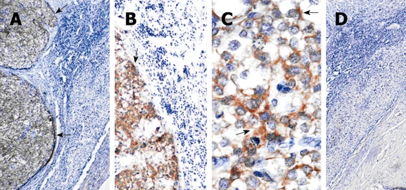
Figure 3.
Immunohistochemistry staining of glypican-3 or α-fetoprotein in liver samples. Representative of the excised hepatocellular carcinoma samples stained with mouse anti-glypican-3 (GPC3) (A-C) or AFP (D, 4 ×); B (10 ×) and c (40 ×) are the amplified image of A (4 ×). The cells stained with yellow or brown particles were considered positive (arrows).

