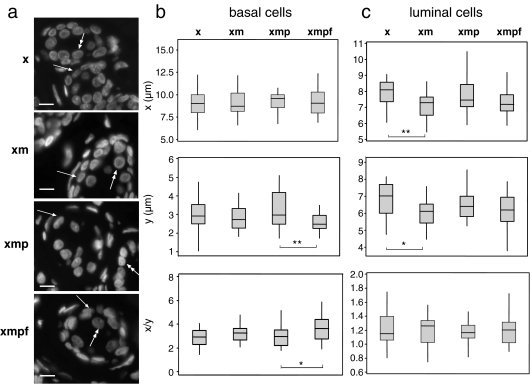
Fig. 2.
Alterations of nuclear size and shape during treatment of breast sections. a DAPI-stained 6-μm breast sections treated with xylene only (x), xylene + microwaving (xm), xylene + microwaving + protease (xmp) and after FISH (xmpf), showing the position of basal (arrow) and luminal (double-headed arrow) epithelial cells. Bars = 10 μm; b box plots of nuclear diameter (micrometres) in x- or y-dimensions and nuclear shape (x/y) for basal epithelial cells in sections treated with xylene only (x), xylene + microwaving (xm), xylene + microwaving + protease (xmp) and as for fish (xmpf). The boxed areas show the 25–75 percentiles and the medians are indicated by horizontal lines through these boxed areas. Statistically significant changes in nuclear size or shape between different treatments are indicated by asterisks (*p < 0.05 and >0.01; **p ≤ 0.01 and >0.001; ***p < 0.001). n = 28; c as in b, but for luminal epithelial cells. n = 28

