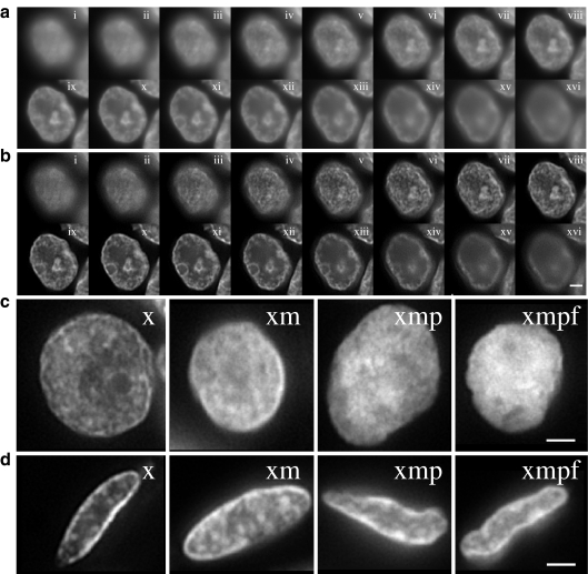
Fig. 3.
Visualisation of chromatin texture during tissue processing. a Grey-scale images of DAPI-stained nucleus from the luminal epithelium of normal mammary gland after xylene treatment to remove wax. Images are taken (i to xvi) at 0.25 μm intervals through the z-axis of the nucleus. Scale bar = 2 μm; b as in a but after image deconvolution to remove out-of-focus information; c single plane deconvolved images of DAPI-stained nuclei from luminal cells of the mammary gland epithelium after treatments with; xylene (x), xylene + microwaving (xm), xylene + microwaving + protease (xmp) and as for fish (xmpf). Scale bar = 2 μm; d as in c but with nuclei from the epithelium of the thyroid gland

