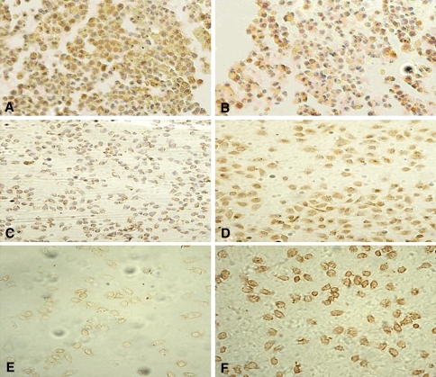
Fig. 4.
Bcl-2, Bax, DR5 protein expression in HepG2 cells (×200). a, c, e Immunocytochemistry of Bcl-2, Bax and DR5 staining in HepG2 cells for 24 h of treatment without DLS. b, d, f Immunocytochemistry of Bcl-2, Bax and DR5 staining in HepG2 cells for 24 h of treatment with 25 μl/ml DLS (All the experiments were repeated four times.)

