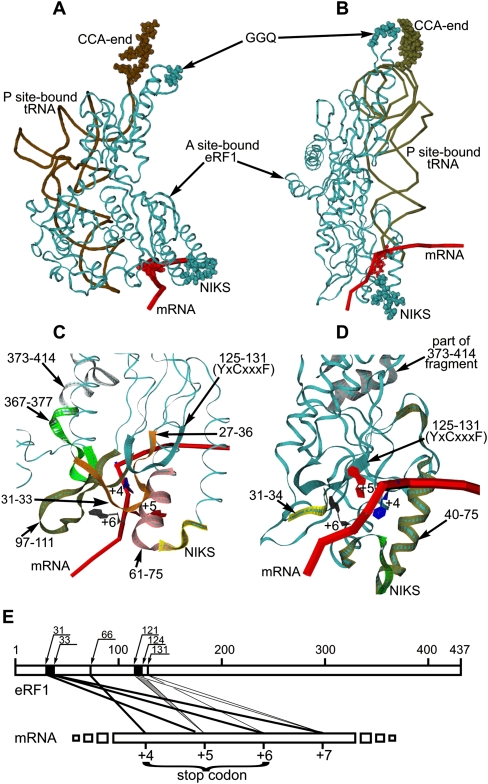
FIGURE 7.
Models M1 (A) and M2 (B) of mutual arrangement of mRNA (red tube corresponding to phosphate backbone; positions +4 are in red beads), the P-site-bound tRNAPhe (brown tube; CCA-ends are in brown beads), and the A-site-bound eRF1_t0 (cyan ribbon; the NIKS and GGQ motifs are in cyan beads). Detailed views of the ribosomal decoding site in models M1 (C) and M2 (D) (mRNA positions +4, +5, and +6 are indicated; the eRF1 motifs given as colored ribbons are indicated by arrows). (E) Schematic representation of the cross-linking sites on eRF1 (numbers on the upper line indicate positions of eRF1 amino acids residues). Major sites are labeled with thick lines.

