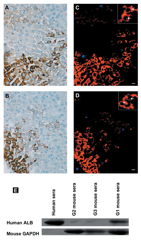
Figure 2.
Engraftment and in vivo differentiation of granulocyte colony-stimulating factor (G-CSF)-mobilized CD34+ HSCs into hepatocyte-like cells in G1 mice 21 days after irradiation and transplantation. Deparaffinized section of liver was stained with (a) anti-human albumin (ALB) antibody or (b) anti-human cytokeratin antibody. (c) Liver was stained with anti-human ALB antibody (red), anti-human CD45 antibody (blue), and human Y chromosome DNA probe (green). (d) Liver was stained with anti-human cytokeratin antibody (red), anti-human CD45 antibody (blue), and human Y chromosome DNA probe (green). The arrow indicates Y chromosome+cytokeratin+ or Y chromosome+ALB+ cells in the liver of G1 mice. The absence of chromosomal staining in some ALB+/cytokeratin+ cell nuclei may be a result of partial sampling of nuclei in the 4-μm thin tissue section. (e) Human serum ALB in the plasma of G1 mice was identified by Western blot analysis. Mouse glyceraldehyde 3-phosphate dehydrogenase (GAPDH) acts as internal standard. Original magnification, (a-d), ×20; inset in (c) and (d), ×60. Representative images are shown.

