
Figure 3. HDACIs improve cell viability in MPP+ treated cells while maintaining histone hyperacetylation.
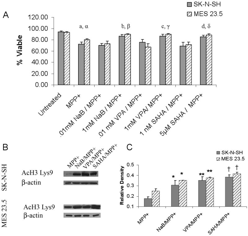
A) Changes in the percent of viable cells estimated by trypan blue exclusion following incubation of cells in MPP+ or MPP+/HDACI. SK-N-SH cells were treated with 100μM MPP+ while MES 23.5 cells were treated with 50μM MPP+. a, α Untreated vs. MPP+ p<0.0001; b, β MPP+ vs. NaB/MPP+ p<0.001; c, γ: MPP+ vs. VPA/MPP+ p<0.001; d, δ: MPP+ vs. SAHA/MPP+ p< 0.01. Incubation of cells with low level HDACIs did not prevent MPP+ mediated decreases in cell viability. B) HDACI-induced hyperacetylation of histone H3 on Lys9 was maintained in the presence of MPP+ (24hr incubation) in both SK-N-SH and MES 23.5 cells. C) Densitometric analysis of acetylated histone H3 Lys9: MPP+ vs. HDACI/MPP+ * p<05; ** p< 0.01; †p< 0.001.