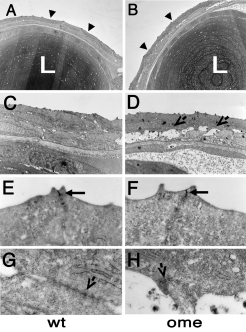
Figure 1.
Electron micrographs of transverse sections through eyes of wild-type and oko meduzy (ome) mutant zebrafish at 3 dpf. (A, B) Low-magnification view of the lens and the overlying cornea. Note that the surface of the mutant cornea features more protrusions than the wild-type cornea. Additionally, fluid-filled spaces are found between epithelial cells in the mutant. Arrowheads: corneal surface. (C, D) Higher-magnification images of the cornea. (D, arrows) Fluid-filled spaces between epithelial cells. (E, F) Higher magnifications of the corneal surface. Wild-type and mutant apical junctions (arrows) do not display any obvious differences. (G, H) Junctions are present on the interface between two layers of corneal epithelial cells. At least some of them persist in the mutant (arrows). L, lens.

