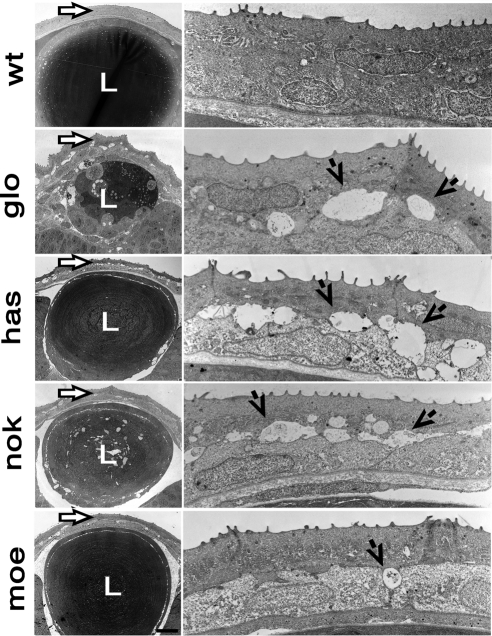
Figure 2.
Electron micrographs of transverse sections through eyes of wild-type and mutant zebrafish at 3 dpf. Left: lens and overlying cornea (arrows). Right: higher magnifications of the corneal epithelium and stromal layer. Each row of images corresponds to a different genotype. Top to bottom: corneas of the wild-type and the following mutant strains: glo, has, nok, and moe. Right, arrows: fluid-filled spaces between epithelial cells of mutant corneas. L, lens.

