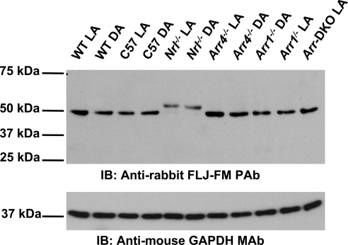
Figure 2.
IB analysis of Als2cr4 protein expression. Each lane contains 20 μg of total retinal lysate from P30 mice. Control WT, Nrl−/−, Arr4−/−, Arr1−/− or Arr-DKO mice were light or dark adapted and killed, and retinal lysates were prepared. The proteins were resolved on 10% SDS-PAGE, transferred onto PVDF membrane, probed with the pAb FLJ-LG followed by the secondary antibody, goat anti-rabbit HRP-conjugated IgG, and processed for ECL. As a loading control, the membrane was probed with the mAb GAPDH and the appropriate secondary antibody.

