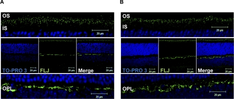
Figure 3.
Als2cr4 immunohistochemical localization. P30 WT mice were either (A) light or (B) dark adapted and killed. The retina sections (7 μm) were incubated with pAb FLJ-FM followed by incubation with Alexa Fluor-488 (Als2cr4) goat anti-rabbit IgG. The nuclei were stained with TO-PRO3 iodide. Top: magnified region of the photoreceptor layer. Middle: view of the retina at ×63 magnification. Bottom: magnified region of the OPL. Results demonstrate a similar staining pattern for Als2cr4 regardless of lighting condition. Punctate staining was observed in the photoreceptor layer, whereas staining in the OPL was more uniform. Magnification, ×63.

