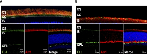
Figure 4.
Dual IHC of Als2cr4 and Arr1 in retinas from light- or dark-adapted mice. P30 WT mice were either (A) light- or (B) dark-adapted and killed. The retina sections were dual stained with mAb D9F2 for Arr1 and pAb FLJ-FM for Als2cr4. Secondary antibodies were Alexa Fluor-568 goat anti-mouse IgG (Arr1) and Alexa Fluor-488 goat anti-rabbit IgG (Als2cr4). Nuclei were stained with TO-PRO3 iodide. (A, top) A magnified region of the merged photoreceptor layer in the bottom panel demonstrates that Als2cr4 was present in the connecting cilia. After 1 hour of light exposure, Arr1 localization was exclusive to the OS, whereas Als2cr4 was present in both the OS and connecting cilia. (B) Al2cr4 was localized in the postsynaptic OPL, with no co-localization of Arr1 with Als2cr4 in rod spherules or cone pedicles. Magnification, ×63.

