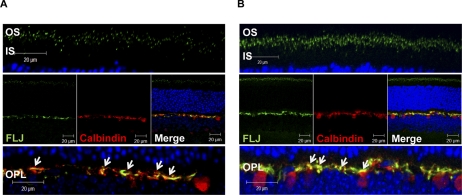
Figure 5.
Dual IHC localization of Als2cr4 and calbindin in horizontal cells. P30 WT mice were either (A) light- or (B) dark-adapted and killed. The retinal sections were dual stained with mAb calbindin and pAb FLJ-FM. Secondary antibodies were Alexa Fluor-568 goat anti-mouse IgG (Calbindin) and Alexa Fluor-488 goat anti-rabbit IgG (Als2cr4). The nuclei were stained with TO-PRO3 iodide. Top: magnified region of the photoreceptor layer. Middle: view of the retina at ×63 magnification. Bottom: magnified region of the OPL. White arrows denote dual immunohistochemical localization of Als2cr4 with calbindin at the OPL. Magnification, ×63.

