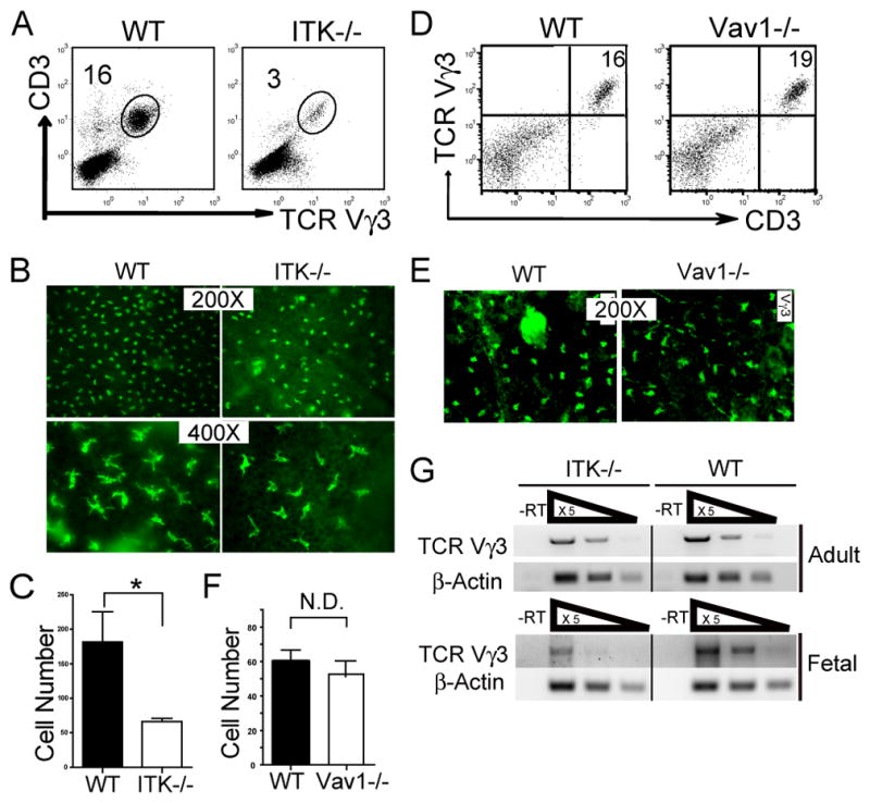
Figure 1.

Impaired development of sIELs in ITK−/− but not Vav1−/− mice. A. Skin cell preparations from 6–8 week old ITK−/− and wild type mice were stained with anti-CD3 and Vγ3 antibodies, and analyzed for percentages of the CD3+Vγ3+ population by flow cytometry. One representative of three independent experiments is shown. B and C. Ear epidermal sheets from wild type and ITK−/− mice were stained with fluorescent anti-Vγ3 antibody and observed under a fluorescent microscope (Olympus BX61) for the Vγ3+ sIELs (B), average numbers of which per field at the 200X amplification were plotted (C). Data were obtained from three independent experiments. *P < 0.05. D-F. Flow cytometric and immunofluorescent microscopic analysis of skin Vγ3+ γδ T cells from Vav1−/− mice as performed in the panels A-C except that the Vγ3+ sIELs on the epidermal sheets was visualized under a different fluorescent microscope (Nikon Eclipse TE 300) that has a smaller field at the same 200X amplification (E). Experiments were repeated twice for both flow cytometric and immunofluorescent analyses. Average numbers of sIELs per field obtained in the panel E were plotted in the panel F. N.D: no difference. G. Total RNA from fetal and adult mouse skin was reverse transcribed to cDNA. Serially 5 fold-diluted cDNA were subject to semi-quantitative PCR to determine expression levels of rearranged TCRγ3 gene. β-actin was used as a control. Data shown were obtained from three independent experiments.
